introducing phase contrast imaging
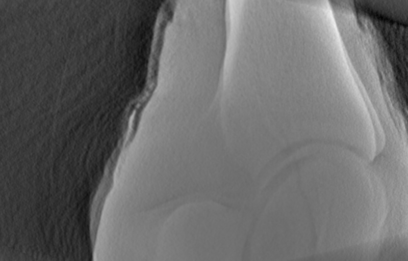
Phase-contrast imaging is known for providing exceptional detail at edges and interfaces in materials. Compare this to absorption imaging where most of this detail is lost.
On the right, the difference between these two techniques is demonstrated. Observe the remarkable changes in detail soft-tissue details.
Use the slider to see the differences!


How it works

Our technology allows you to retain the traditional X-ray image, get two new X-ray images with enhanced detail for diagnosis without significant disruption to existing diagnostic processes.
New Hardware:
-
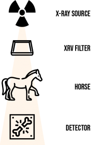
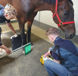
- Filter – Single component, retro-fitted to existing machines (CT or planar X-ray machines)
New Images:
-
- X-ray – Retain the original X-ray image
- XR EBI – Enhanced Bone Image (CT-like features)
- XR STS – Soft Tissue Scan (MRI-like features)

Imaging workflow
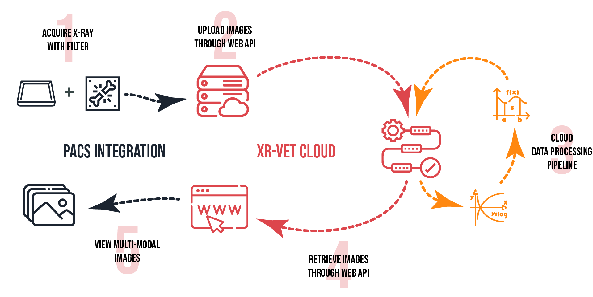
Our technology is designed to integrate with PACS systems. Simply acquire an image using our filter on your existing X-ray system, and pass the image through our cloud processing pipeline and instantly receive the conventional X-ray plus two new images unique to XR-Vet: an Enhanced Bone Image (EBI) and Soft Tissue Scan (STS).

